
Dnipro State Medical University
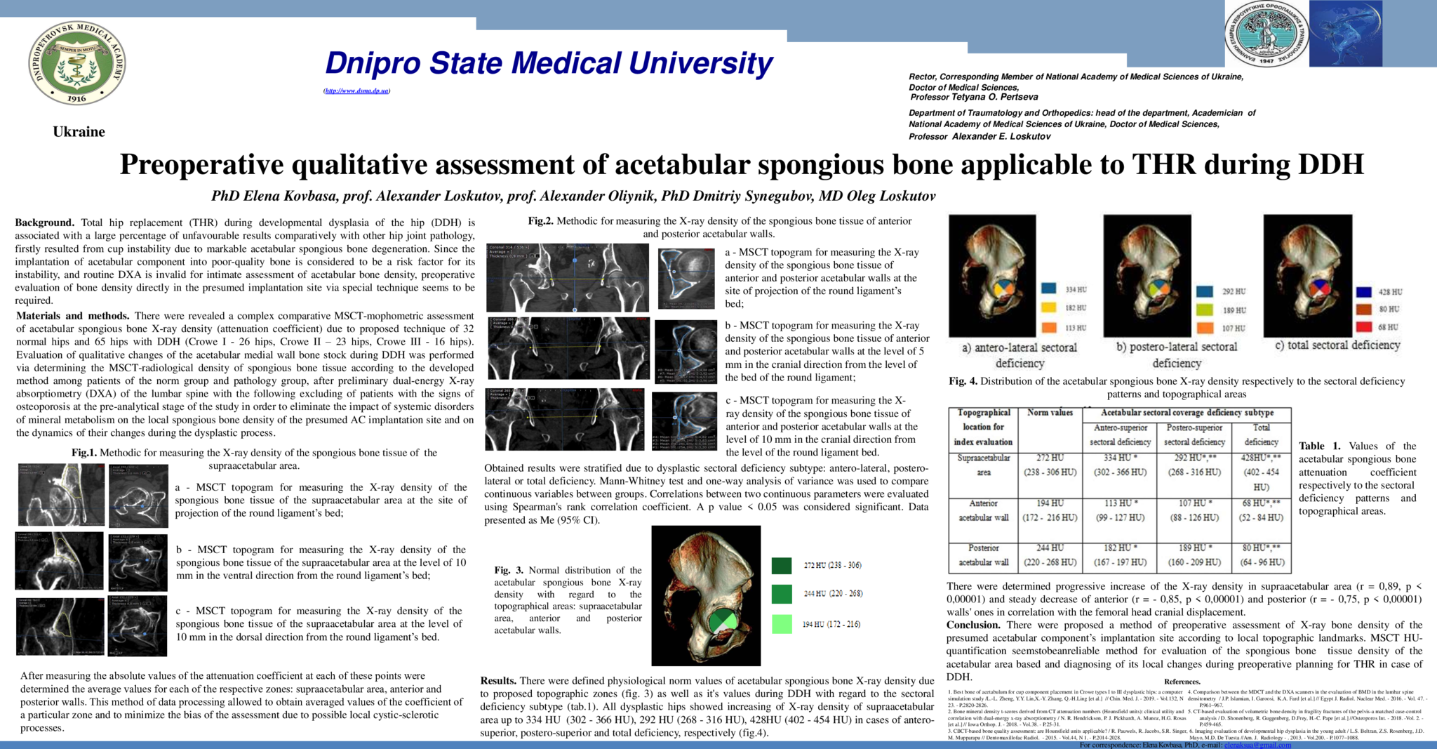
INTRODUCTION: Since the implantation of the acetabular component into poor-quality bone is considered to be a risk factor for its instability, and routine DXA is invalid for intimate assessment of acetabular bone density, preoperative evaluation of bone density directly in the presumed implantation site via special method seems to be required. MATERIALS AND METHODS: There were revealed a complex comparative MSCT-mophometric assessement of acetabular spongious bone X-ray density (attenuation coefficient) due to proposed methodic of 32 normal hips and 65 hips with DDH (Crowe I - 26 hips, Crowe II - 23 hips, Crowe III - 16 hips). Patients with DXA-verified osteoporosis or osteopenia were excluded. The evaluation implied 5 mm-interval measuring due to topographical zones (supraacetabular area, anterior and posterior acetabular walls). Obtained results were stratified due to dysplastic sectoral deficiency subtype. Mann-Whitney test and one-way analysis of variance was used to compare continuous variables between groups. Correlations between two continuous parameters were evaluated using Spearman's rank correlation coefficient. A p value < 0.05 was considered significant. Data presented as Me (95% CI). RESULTS: There were defined physiological norm values (fig. 1) of acetabular spongious bone X-ray density due to proposed topographic zones, as well as it's values during different sectoral deficiency subtypes (table 1). There were determined progressive increase of the X-ray density of the supraacetabular area (r = 0,89, p < 0,001) and steady decrease of anterior (r = - 0,85, p < 0,001) and posterior (r = - 0,75, p < 0,001) walls ones in correlation with the femoral head cranial displacement. The most severe loss of acetabular spongious bone X-ray density in all the zones was observed during the total sectoral deficiency subtype. CONCLUSION: Proposed method of evaluation and conducted regularities should be taken into account for preoperatite planing of AC implantation during DDH.
- 69 προβολές
Abstract ID
ΑΑ029