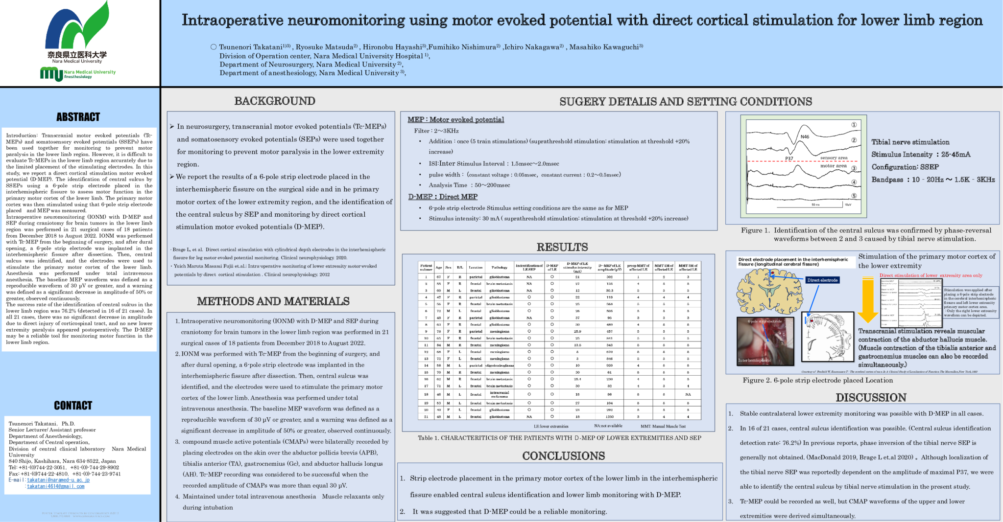
Introduction: Transcranial motor evoked potentials (Tc-MEPs) and somatosensory evoked potentials (SSEPs) have been used together for monitoring to prevent motor paralysis in the lower limb region. However, it is difficult to evaluate Tc-MEPs in the lower limb region accurately due to the limited placement of the stimulating electrodes. In this study, we report a direct cortical stimulation motor evoked potential (D-MEP). The identification of central sulcus by SSEPs using a 6-pole strip electrode placed in the interhemispheric fissure to assess motor function in the primary motor cortex of the lower limb. The primary motor cortex was then stimulated using that 6-pole strip electrode placed and MEP was measured. Intraoperative neuromonitoring (IONM) with D-MEP and SEP during craniotomy for brain tumors in the lower limb region was performed in 21 surgical cases of 18 patients from December 2018 to August 2022. IONM was performed with Tc-MEP from the beginning of surgery, and after dural opening, a 6-pole strip electrode was implanted in the interhemispheric fissure after dissection. Then, central sulcus was identified, and the electrodes were used to stimulate the primary motor cortex of the lower limb. Anesthesia was performed under total intravenous anesthesia. The baseline MEP waveform was defined as a reproducible waveform of 30 μV or greater, and a warning was defined as a significant decrease in amplitude of 50% or greater, observed continuously. The success rate of the identification of central sulcus in the lower extremity limb region was 71.4% (detected in 15 of 21 cases). In all 21 cases, there was no significant decrease in amplitude due to direct injury of corticospinal tract, and no new lower extremity paralysis appeared postoperatively. The D-MEP may be a reliable tool for monitoring motor function in the lower limb region.
- 1 view


