National & Kapodistrian University of Athens, School of Medicine, 4th Department of Surgery, ATTIKON University Hospital
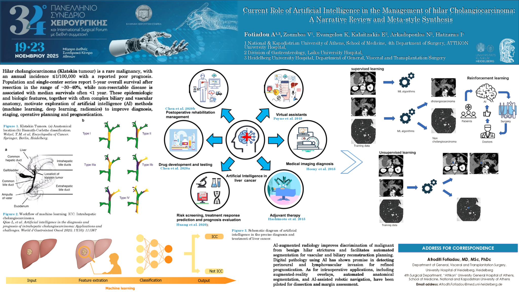
Introduction: Hilar cholangiocarcinoma (Klatskin tumour) is a rare malignancy, with an annual incidence ≲1/100,000 with a reported poor prognosis. Population and single-center series report 5-year overall survival after resection in the range of ~30–40%, while non-resectable disease is associated with median survivals often <1 year. These epidemiologic and biologic features, together with often complex biliary and vascular anatomy, motivate exploration of artificial intelligence (AI) methods (machine learning, deep learning, radiomics) to improve diagnosis, staging, operative planning and prognostication.
Aim: The aim of this narrative review is to synthesize current evidence on AI applications relevant to the multidisciplinary management of Klatskin tumours, with emphasis on studies applicable to hepatobiliary surgery.
Material and Method: We performed a narrative PubMed/MEDLINE review (last search September 2025) combining terms for hilar cholangiocarcinoma/Klatskin tumour and AI across imaging, pathology, multi-omics and intraoperative domains. Clinical studies, systematic reviews and meta-analyses reporting diagnostic, staging, prognostic or management-relevant endpoints were prioritized; evidence was synthesized narratively and pooled metrics reported when available.
Results: AI-augmented radiology improves discrimination of malignant from benign hilar strictures and facilitates automated segmentation for vascular and biliary reconstruction planning. In a cohort of 274 patients, Quin et al. reported a multilevel radiomics–machine learning model predicting early recurrence with an AUC of 0.883, outperforming conventional staging. Similarly, Zhan et al. developed a CT-radiomics model for lymph node metastasis (n=214), achieving AUCs of 0.98 (training) and 0.87 (external validation), and stratifying survival (median 13.7 vs 27.3 months). Digital pathology using AI has shown promise in detecting perineural and lymphovascular invasion for refined prognostication. As for intraoperative applications, including augmented-reality overlays, automated anatomical segmentation, and AI-assisted robotic navigation, have been piloted for dissection and margin assessment. A single-center robotic series of 21 Klatskin patients achieved R0 resection in 90% with median blood loss of 150 mL and operative time of 458 minutes. A meta-analysis of 267 patients comparing robotic and open surgery found no significant differences in operative time, blood transfusion, R0 rates, or complications, supporting feasibility.
Conclusions: AI is emerging as a valuable adjunct in imaging, pathology, and surgical planning for Klatskin tumours, with immediate utility in preoperative staging and intraoperative guidance. Translation into routine practice requires multi-institutional datasets, prospective validation, and demonstration of patient-level benefits such as improved resectability, margin status, and survival
- 4 προβολές
Abstract ID
ΑΑ294
Συγγραφέας

Fotiadou Afroditi
Resident Surgeon
Department of General, Visceral and Transplantation Surgery, Heidelberg University Hospital
- 4 προβολές