Theagenio Cancer Hospital Thessaloniki Greece, Hellenic Society of Medical Oncology
Introduction: DJ-1 is involved in various cellular processes. An increasing number of studies have found that the amount of DJ-1 is overexpressed in most known human cancers, which is consistent with its role as an oncogene. Interestingly, DJ-1 could be secreted by cancer cells and thus could serve as a biomarker for cancer monitoring.
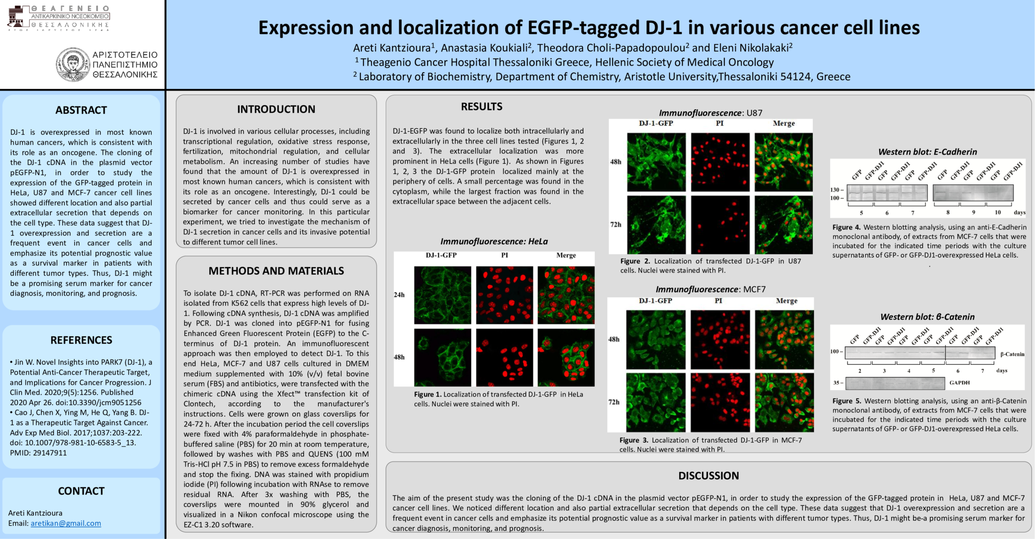
Methods: As a first step to monitor the expression and localization of DJ-1 both in the various subcellular compartments but also extracellularly, we used Green Fluorescent Protein as a DJ-1 tag and performed immunofluorescence experiments.
To isolate DJ-1 cDNA, RT-PCR was performed on RNA isolated from K562 cells that express high levels of DJ-1. cDNA was isolated from recombinant pGEM®-T Easy and ligated into the EcoRI/ BamHI site of pEGFP-N1 for fusing Enhanced Green Fluorescent Protein (EGFP) to the C-terminus of DJ-1 protein.
An immunofluorescent approach was then employed to determine DJ-1 which was tagged at its C terminus with EGFP. To this end HeLa, MCF-7 and U87 cells cultured and antibiotics were transfected with the chimeric cDNA using the Xfect™ transfection kit of Clontech, according to the manufacturer's instructions. Cells were grown on glass coverslips for 24-72 h. After 3x washing with PBS, the coverslips were mounted in 90% glycerol and visualized in a Nikon confocal microscope .
Results: DJ-1-EGFP was found to localize both intracellularly and extracellularly in the three cell lines tested .The extracellular localization was more prominent in HeLa cells The DJ-1-GFP protein localized mainly at the periphery of cells.
Discussion: The aim of the present study was the cloning of the DJ-1 cDNA in the plasmid vector pEGFP-N1, in order to study the expression of the GFP-tagged protein in HeLa, U87 and MCF-7 cancer cell lines. We noticed different location and also partial extracellular secretion that depends on the cell type.
- 10 προβολές
Abstract ID
AA-21