Introduction: Masson’s lesion or also known as Intravascular papillary endothelial hyperplasia is a rare form of reactive endothelial proliferation which can mimic benign and malignant vascular tumors. It commonly occurs on the head, neck, trunk, and upper extremities, but rarely in the foot. It is important to include it in the differential diagnosis of malignant vascular neoplasms, such as angiosarcoma, in order to avoid aggressive treatment.
Aim: We present a case of this type of tumor at the lateral region of the foot. Our aim is to describe the pathogenic and histologic features of this lesion.
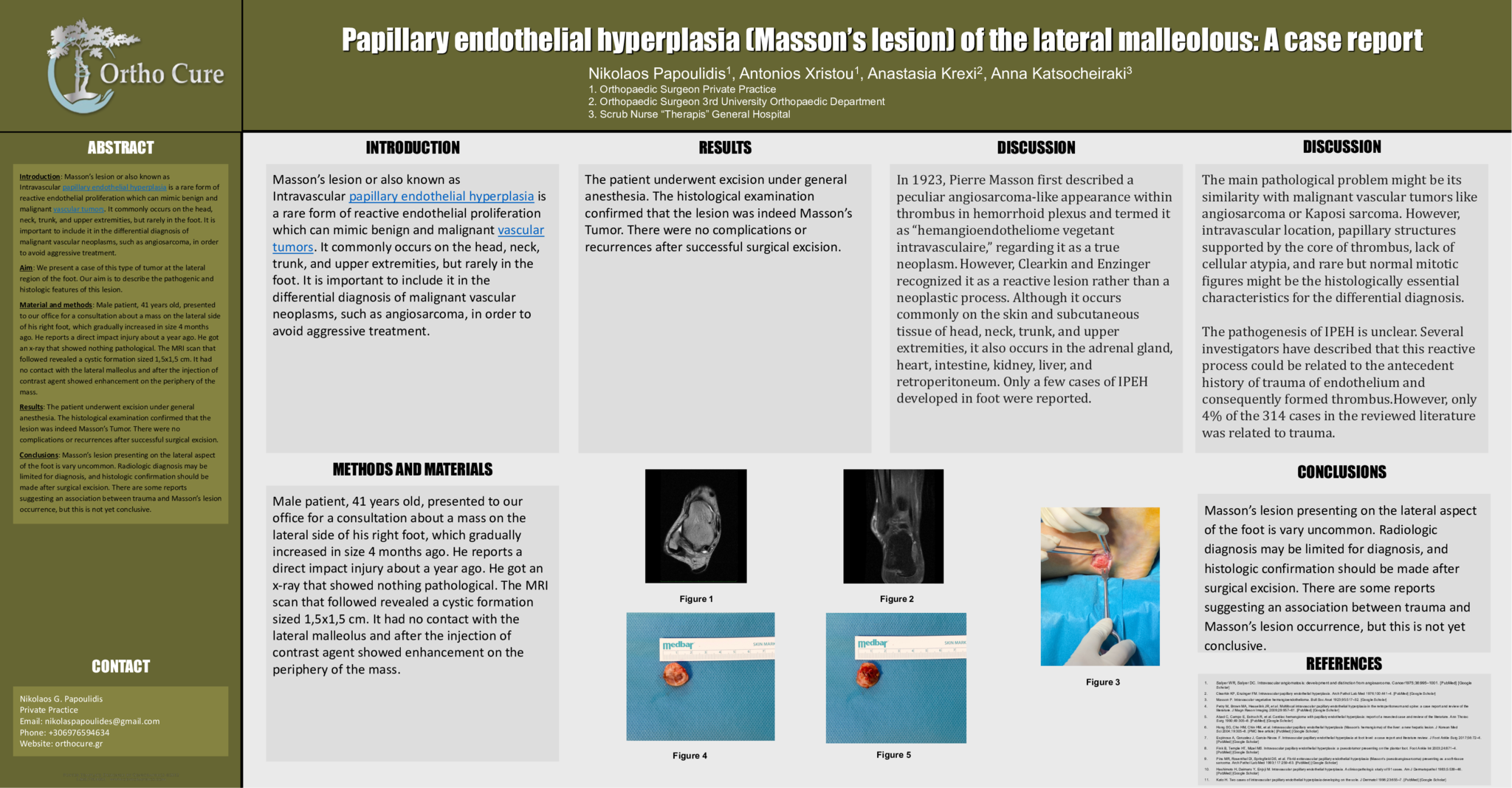
Material and methods: Male patient, 41 years old, presented to our office for a consultation about a mass on the lateral side of his right foot, which gradually increased in size 4 months ago. He reports a direct impact injury about a year ago. He got an x-ray that showed nothing pathological. The MRI scan that followed revealed a cystic formation sized 1,5x1,5 cm. It had no contact with the lateral malleolus and after the injection of contrast agent showed enhancement on the periphery of the mass.
Results: The patient underwent excision under general anesthesia. The histological examination confirmed that the lesion was indeed Masson’s Tumor. There were no complications or recurrences after successful surgical excision.
Conclusions: Masson’s lesion presenting on the lateral aspect of the foot is vary uncommon. Radiologic diagnosis may be limited for diagnosis, and histologic confirmation should be made after surgical excision. There are some reports suggesting an association between trauma and Masson’s lesion occurrence, but this is not yet conclusive.
- 6 προβολές


