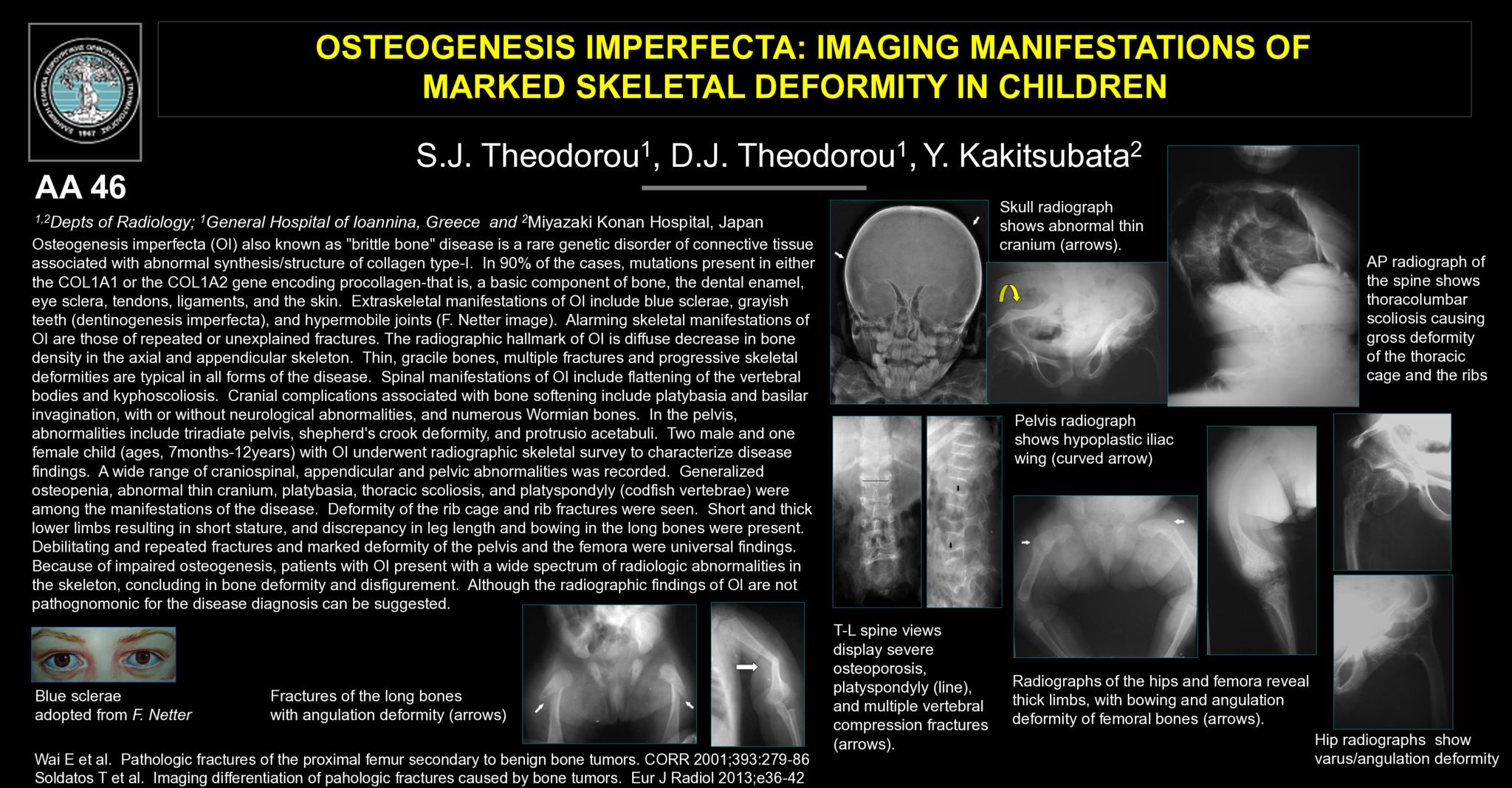
Background: Osteogenesis imperfecta (OI) also known as "brittle bone" disease is a rare genetic disorder of connective tissue associated with abnormal synthesis/structure of collagen type-I. In 90% of the cases, mutations present in either the COL1A1 or the COL1A2 gene encoding procollagen-that is, a basic component of bone, dental enamel, eye sclera, tendons, ligaments, and the skin. Extraskeletal manifestations of OI include blue sclerae, grayish teeth (dentinogenesis imperfecta), and hypermobile joints. Alarming skeletal manifestations of OI are those of repeated or unexplained fractures. The radiographic hallmark of OI is diffuse decrease in bone density in the axial and appendicular skeleton. Thin, gracile bones, multiple fractures and progressive skeletal deformities are typical in all forms of the disease. Spinal manifestations of OI include flattening of the vertebral bodies and kyphoscoliosis. Cranial complications associated with bone softening include platybasia and basilar invagination, with or without neurological abnormalities, and numerous Wormian bones. In the pelvis, abnormalities include triradiate pelvis, shepherd's crook deformity, and protrusio acetabuli.
Purpose: Two male and one female child (ages, 7months-12years) with OI underwent radiographic skeletal survey to characterize disease findings.
Findings: A wide range of craniospinal, appendicular and pelvic abnormalities was recorded. Generalized osteopenia, abnormal thin cranium, platybasia, thoracic scoliosis, and platyspondyly (codfish vertebrae) were among the manifestations of disease. Deformity of the rib cage and rib fractures were seen. Short and thick lower limbs resulting in short stature, and discrepancy in leg length and bowing in the long bones were present. Debilitating and repeated fractures and marked deformity of the pelvis and the femora were universal findings.
Summary: Because of impaired osteogenesis, patients with OI present with a wide spectrum of radiologic abnormalities in the skeleton, concluding in bone deformity and disfigurement. Although the radiographic findings of OI are not pathognomonic for the disease diagnosis can be suggested.
- 1 προβολή


