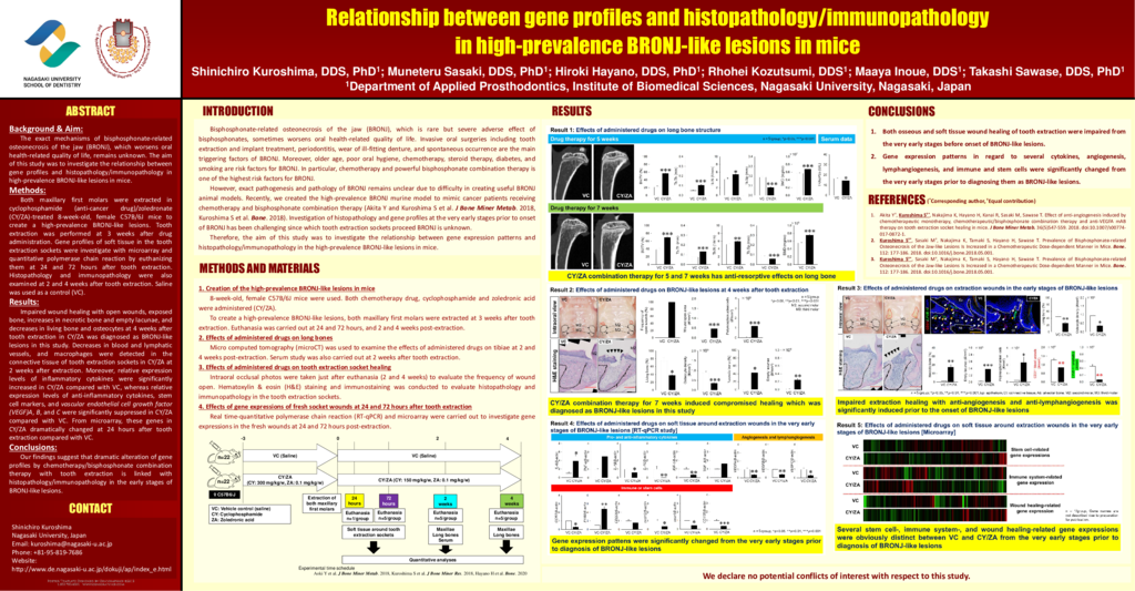
Background & Aim: The exact mechanisms of bisphosphonate-related osteonecrosis of the jaw (BRONJ), which worsens oral health-related quality of life, remains unknown. The aim of this study was to investigate the relationship between gene profiles and histopathology/immunopathology in high-prevalence BRONJ-like lesions in mice. Methods: Both maxillary first molars were extracted in cyclophosphamide (anti-cancer drug)/zoledronate (CY/ZA)-treated 8-week-old, female C57B/6J mice to create a high-prevalence BRONJ-like lesions. Tooth extraction was performed at 3 weeks after drug administration. Gene profiles of soft tissue in the tooth extraction sockets were investigate with microarray and quantitative polymerase chain reaction by euthanizing them at 24 and 72 hours after tooth extraction. Histopathology and immunopathology were also examined at 2 and 4 weeks after tooth extraction. Saline was used as a control (VC). Results: Impaired wound healing with open wounds, exposed bone, increases in necrotic bone and empty lacunae, and decreases in living bone and osteocytes at 4 weeks after tooth extraction in CY/ZA was diagnosed as BRONJ-like lesions in this study. Decreases in blood and lymphatic vessels, and macrophages were detected in the connective tissue of tooth extraction sockets in CY/ZA at 2 weeks after extraction. Moreover, relative expression levels of inflammatory cytokines were significantly increased in CY/ZA compared with VC, whereas relative expression levels of anti-inflammatory cytokines, stem cell markers, and vascular endothelial cell growth factor (VEGF)A, B, and C were significantly suppressed in CY/ZA compared with VC. From microarray, these genes in CY/ZA dramatically changed at 24 hours after tooth extraction compared with VC. Conclusions: Our findings suggest that dramatic alteration of gene profiles by chemotherapy/bisphosphonate combination therapy with tooth extraction is linked with histopathology/immunopathology in the early stages of BRONJ-like lesions.
- 41 views


